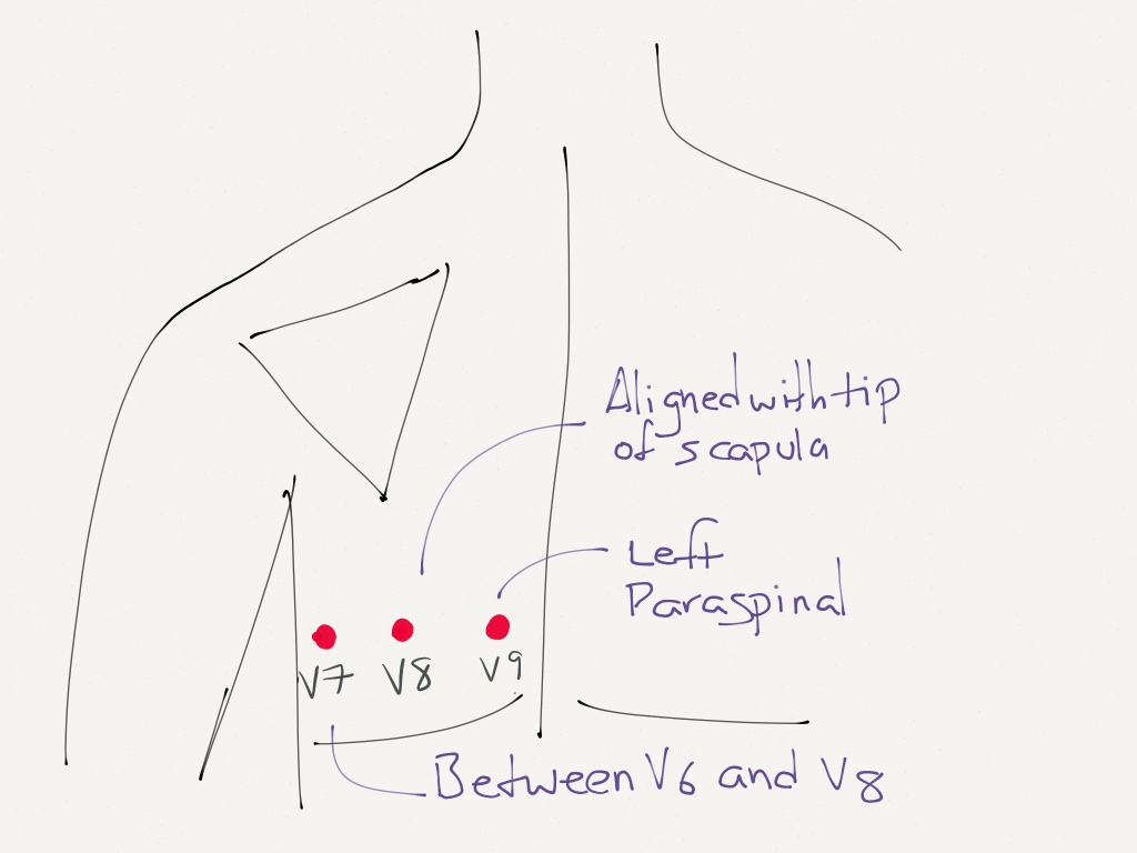
Remeber your coronary artery anatomy. V7 Left posterior axillary line in the same horizontal plane as V6.
Lead Placement For Posterior Ecg Resus Review
Click to read in-depth answer.
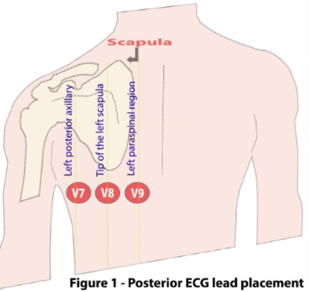
. V8 Tip of the left scapula in the same horizontal plane as V6. ELECTROCARDIOGRAM ALTERNATE LEAD PLACEMENTS RIGHT SIDED OR V7 V8 V9 2140712 Procedure Posterior V 7-9 ECG 1 Perform a routine 12 lead ECG with regular limb and chest lead placement. Posterior MIs often co-exist with inferior or lateral STEMI.
The editor and publisher of this work have checked with sources believed to be reliable in their efforts to provide. V8 Tip of the left scapula in the same horizontal plane as V6. Inferior angle of the scapula.
Left posterior axillary line V8. V7 is placed at the posterior axillary line in the same horizontal plane as V6. It is also helpful for future clinicians if you note in your read that it is a posterior ECG.
V9 or c 9 is placed along the left spinal border at the. To clarify leads will equal. Posterior leads Leads V7-9 are placed on the posterior chest wall in the following positions see diagram below.
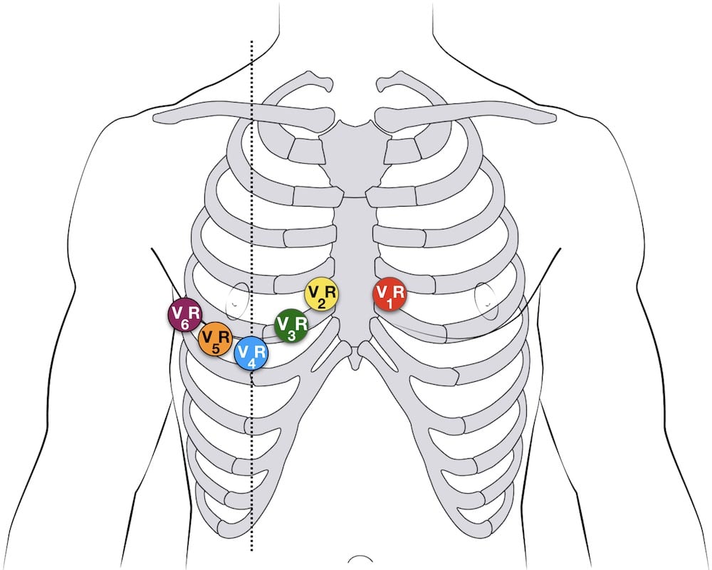
V9 same horizontal line as V4R left paraspinal border use V6 electrode. 4 right chest leads denoted by V3R V4R V5R and V6R. V8 is placed at the tip of the left scapula in the same horizontal plane.
At a minimum lead V4 should be placed on the 5th intercostal mid-clavicular exact opposite of the regular left side placement if an inferior infarct was originally seen in leads II III and AVF. Level with V7 at mid-scapular line V9. Nonetheless for the SDB pairs v2 -v3 and v7 -v8 the trained model achieved both good precision and recall.
V7 is located at the same horizontal line as V4R ie 5th ICS on the posterior axillary line use the V4 electrode. Once the electrocardiogram with posterior leads has been made you must write the word Posteriors in the EKG header and overwrite leads V7 V8 V9 on the leads that have been replaced by posterior leads. V9 is placed in the left paraspinal region in the same horizontal plane.
The rock of Powerlinez can easily be seen when driving north on the New York Thruway just a couple miles north from the big interchange of routes 87 287 17 at the southern border of NY state with New Jersey. At the same level as electrode V6 and the midscapular line tip of the scapula. V8 same horizontal line as V4R mid subscapular line use V5 electrode.
V7 Left posterior axillary line in the same horizontal plane as V6. V4V7 V5V8 and V6V9. Placement of Posterior Leads.
V9 is placed in the left paraspinal region in the same horizontal plane. Leads V7-V8-V9 can be used to diagnose a posterior infarct. Left tip of scapula V9.
V8 is placed at the tip of the left scapula in the same horizontal plane. Leads V7-9 are placed on the posterior chest wall in the following positions see diagram below. V7 is placed at the posterior axillary line in the same horizontal plane as V6.
Placement of posterior leads V7-V9. Move V4 V5 V6 to posterior positions V7. Left paraspinal region Look for ST elevations in V7 V8 V9 on your p osterior EKG.
See the chapter Ischemia for other ways of diagnosing posterior infarction. At the same level as electrodes V6 the left paravertebral line. Level with V6 at left posterior axillary line V8.
Leads V7-9 are placed on the posterior chest wall in the following positions. V8 or c8 is placed in the left mid scapular line at the same horizontal level as v4 v. On most EKg machines the labels areno automatically changed so it is important to cross out the labels for V4-V6 and write in V7-V9.
Level with V8 just left of vertebral line Special Lead Placement. V9 Left paraspinal region in the same horizontal plane as V6. What is the correct placement of leads V7 V9.
Posterior Ventricular leads V7 V8 V9. V9 Left paraspinal region in the same horizontal plane as V6. Lastly a right sided 12-lead ECG placement allows you to detect a right sided infarct.
In some clinical situations the recording of posterior electrode positions may be necessary posterior electrodes are placed in the same transverse plane as v4. Pick up V4 V5 V6 and replace with V7 V8 V9 V7. V9 Left paraspinal region in the same horizontal plane as V6.
FAACN Essentials of Critical Care NursingPocket Handbook f Notice Medicine is an ever-changing science. V7 or c7 is placed in the left posterior axillary line at the same horizontal level as v4. V7 Left posterior axillary line in the same horizontal plane as V6.
Leads to improve detection of atrial rhythm. After V6 leads are placed towards the back. The performance variations imply that for certain versions it is difficult to differentiate between dependencies and non-dependencies FT due to similar structural characteristics.
2 Reposition the chest electrodes per the attached diagram for V 7 V 8 V 9 on the patients back. V8 Tip of the left scapula in the same horizontal plane as V6. These include the Frank vectorcardiogram VCG system which uses 3 nearly orthogonal leads denoted as X Y and Z.
The largest rock climbing area within an hours drive from New York City -- in the southwest corner of Harriman State Park. Lead Placement for Posterior ECG Resus Review. The leads V4-V6 are removed and substituted for V7-V9 as shown below.
V4R V4 but right sided is a sensitive lead for diagnosing right ventricular infarctions. Lead Placement for Posterior ECG. This blog aims to disrupt how medical providers and trainees can gain public access to high-quality educational content while also engaging in a dialogue about best-practices in EM and medical education.
Just to the lateral to the vertebrae. As new research and clinical experience broaden our knowledge changes in treatment and drug therapy are required. And 3 left posterior leads denoted as V7 V8 and V9.
Stemi Equivalents Maimonides Emergency Medicine Residency

Ecg Lead Positioning Litfl Ecg Library Basics

Posterior Electrode Placement V7 Is Placed In The Left Posterior Download Scientific Diagram

Electrocardiographic Diagnosis Of Remote Posterior Wall Myocardial Infarction Using Unipolar Posterior Lead V9 Chest

How To Not Miss A Posterior Myocardial Infarction Em Daily

Diagnostics Alternative Ekg Leads Taming The Sru
Ecg Lead Positioning Litfl Ecg Library Basics

Active Chest Pain Trop 5 0 Core Im Podcast
0 comments
Post a Comment